Table of Contents
Approved
Over the past few days, some of our users have encountered a known error where the tumor was not detected on ultrasound or mammography. This problem is caused by many factors. Let’s find out about them below. An ultrasound or mammogram is often used to inform the surgeon about an abnormal segment of the breast. If your breast is not clearly identified in any exam, it will still need to be examined and a biopsy will likely be needed to determine if it is cancerous or not.
An ultrasound or mammogram is often used to guide the surgeon to an abnormal area of your breasts. If a lump in your breast is not visible in any study, it still needs to be examined and a large biopsy will likely be needed to determine if it qualifies as cancer.
Certain cancers can be missed on a mammogram. Some breast changes, including the lumps you feel, will not show up on a mammogram. One woman said that although her first mammogram was crystal clear, four months later, she discovered any cancers.
A pectoral bump is a bump that grows in relation to the chest. Breast lumps vary in size and texture and can be painful. Some of them end up not being found on physical imaging or examination. Most breast growths are benign (not cancerous).
Your specialist will most likely perform a physical exam to assess breast lumpiness. To determine if a tumor is cancerous, your doctor will likely order a mammogram and an ultrasound of your breasts.What type. In addition, MRI, PET/CT of the breast, or scintimography may be performed. If the tumor is confirmed to be benign, further action may be needed, but your doctor may want to examine it to see if it changes, grows, or disappears over time. If test results are inconclusive, an absolute biopsy using ultrasound, X-ray, or permanent resonance imaging may be done. If my tumor is confirmed to be cancerous, surgery is usually performed. Additional treatment may include therapy, radiation therapy, or hormone therapy.
Inflammatory breast cancer (IBC) differs from other types of breast cancer for several reasons: IBC is not like recurrent breast cancer. This often does not cause lumps to form in the breast, and this type may not appear on a real mammogram.
This page was modified on January 31, 2020.
Which Breast Is Tight?
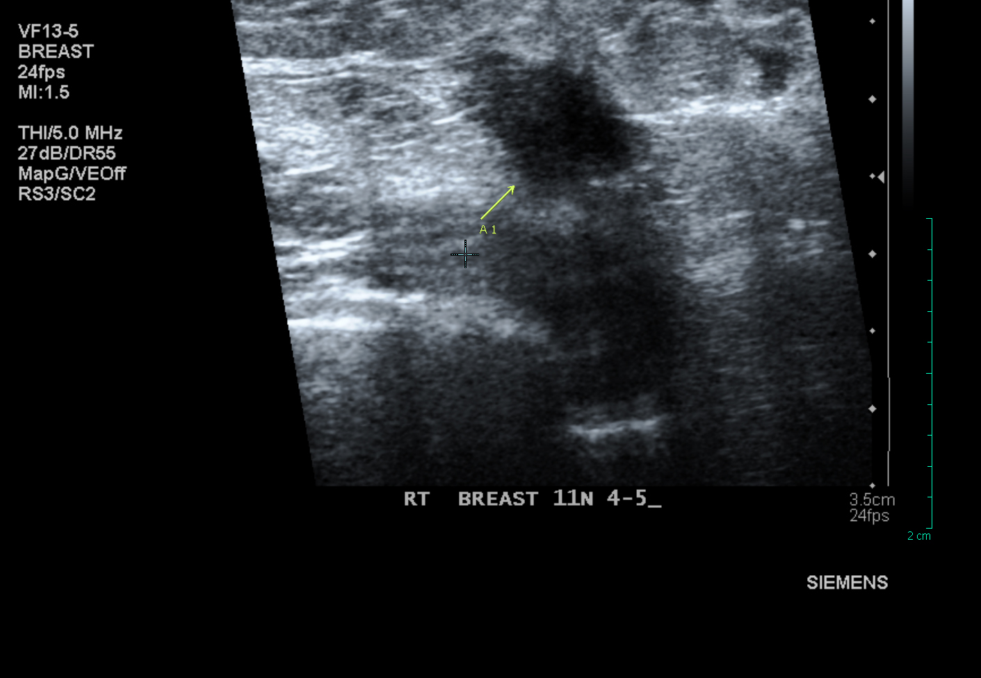
Indeed, this remedy may miss some of the early signs of cancer. An example of the first clues that may not appear on ultrasound are small deposits that are considered microcalcifications. Ultrasound can be used if: you have particularly dense tissue in the nipple area.
Approved
The ASR Pro repair tool is the solution for a Windows PC that's running slowly, has registry issues, or is infected with malware. This powerful and easy-to-use tool can quickly diagnose and fix your PC, increasing performance, optimizing memory, and improving security in the process. Don't suffer from a sluggish computer any longer - try ASR Pro today!

A breast tumor is a tumor that is constantly growing in the breast. Depending on the type of lump in the nipple area may be large, small, hard or soft to the touch. Some nodules cause pain, while weak nodules go unnoticed until they are detected with a high-quality imaging study.
The seal can beb Discovered by a woman doing a breast self-exam or by a doctor during a physical examination. Suspicious lumps can also be found during an annual mammogram screening. Although rare, hemorrhoids can occur in men.
It’s helpful to be familiar with the normal look and feel of your own breasts, and where they feel, so you can let your doctor know about any changes.
How Are Breast Masses Diagnosed And Evaluated?
Most breast lumps are
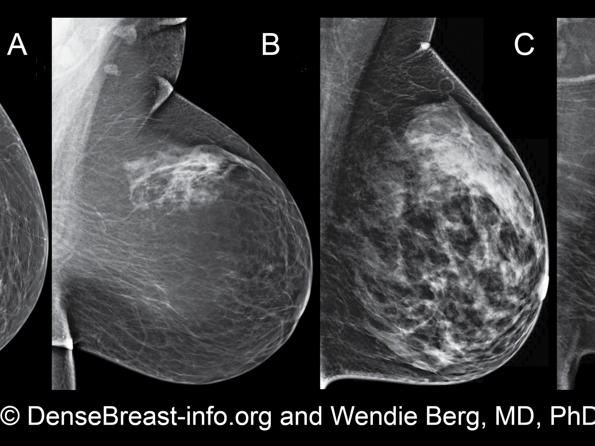
About 20% to 30% of young women with breast cancer have tumors that are not visible on mammograms. And these interval breast cancers found between normal mammograms seem to be more lethal than those found.at screening.
If a tumor that appears to be benign on these examinations turns out to be benign, no further action should be taken. Your doctor should monitor the area during follow-up visits to check if the breast swelling has changed or gone.
Indeed, some early signs of cancer may go unnoticed. An example of early signs that cannot be explained by ultrasound are tiny deposits of calcium minerals called microcalcifications. Ultrasound can be used continuously if you have: especially shiny breast tissue.
If these tests do not conclusively indicate that the tumor is benign,
 The software to fix your PC is just a click away - download it now.
Approximately 20-30 percent of women with breast cancer develop tumors that go unnoticed during mammography screening. And these interval cancersbreast cancers found between routine mammograms appear to be more lethal than those found on screening.
Mammography can miss some types of cancer. Some breast changes, including palpable hemorrhoids, cannot be seen on a mammogram. A loved one says that although her number one mammogram was clear, four months later, the author discovered a tumor while playing, which turned out to be cancer.
The sensitivity and then the specificity of US for the detection of breast enlargement carcinoma were 57.1% and 62.8%, respectively, with a positive predictive value of 68.1%, a negative predictive value of 99.5%, a positive odds ratio of 39, and a negative one. odds ratio 0.07.
![]()
![]()
![]()
![]()
![]()
![]()
![]()
![]()
![]()
![]()